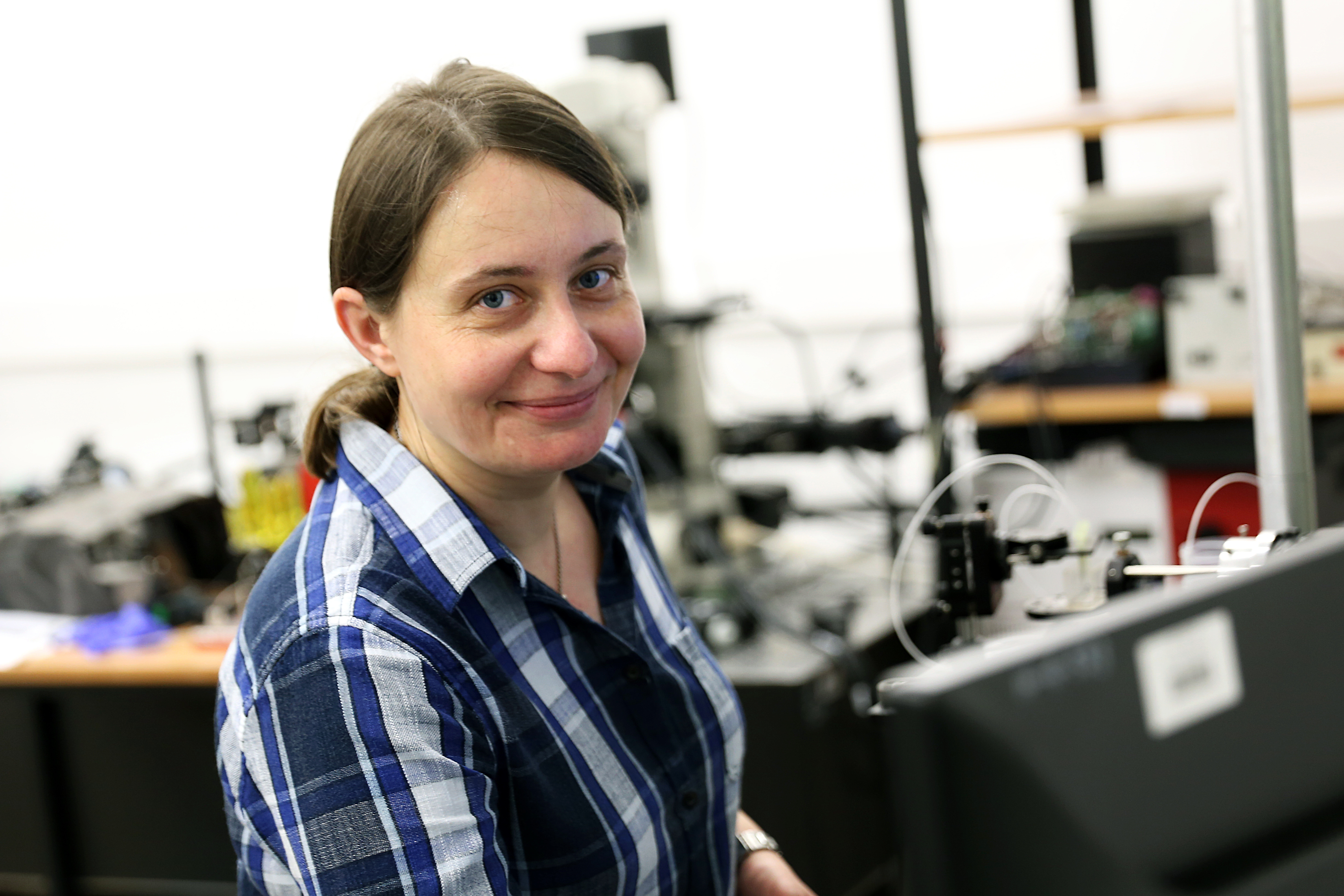
Iwona Gorczynska, PhD, is an assistant professor at Faculty of Physics, Nicolaus Copernicus University, Torun, Poland. Her research focuses on development of biomedical imaging methods utilizing optical coherence tomography techniques, with applications in ophthalmology and microscopy. Dr Gorczynska has received her doctoral degree from Department of Physics, Astronomy and Informatics, Nicolaus Copernicus University in Torun, Poland. She has completed a scientific training at University of Kent at Canterbury, United Kingdom as a Marie Currie Training Site fellow. She did her postdoctoral training at the Optics and Quantum Electronics Group at Research Laboratory of Electronics, MIT, Cambridge, MA, USA and worked as a visiting assistant professor at Vision Science and Advanced Retinal Imaging Laboratory, University of California Davis, Sacramento, CA, USA.
How to look deep into the eyes…
…and study the structure and function of the retina and ocular vascular systems? This is the question that lies at the foundation of the research which I have started during my doctoral studies at the Faculty of Physics, Astronomy and Applied Informatics. I continue researching different aspects of imaging of the living tissue ever since. The technique which I exploit is optical coherence tomography (OCT).
OCT enables three-dimensional imaging of objects partly transparent to light, and may be used for in vivo and non-invasive imaging of biological tissues. The carrier of information about the studied object is light. It is emitted by specially-designed laser sources and formed into a beam which penetrates harmlessly into the object. When the beam propagates inside an object, it encounters a variety layers with different optical properties. These layers may transform some parameters of the light, for example its intensity or frequency (due to dispersion or absorption of the optical medium). Some of the light back-scattered in the subsequent layers returns towards the detector, carrying information about the structure, and functional processes occurring in the object. The information about volumetric distribution of the tissue parameters of interest is obtained by analysis of the signal registered at different time delays, corresponding to light scattered at different depths of the object. However, a direct analysis of those delays is not possible, since the speed of light is nearly 300 thousand km per second, and travels the distance of 1 mm within biological tissues in ~5 picoseconds (1 picosecond is one trillionth part of a second). At present there are no detectors fast enough to allow for direct measurement of such short periods of time. Instead, a ‘trick’ is used, which was devised as early as in the 19th century by Albert Michelson (a Nobel Prize winner born in Strzelno near Toruń), and known to physicists as “white light interferometry”. It involves splitting a beam of light into two parts. One reaches a reference mirror, and the other penetrates into the object. Both beams return towards the detector, at which they are superimposed creating so called interference signal – a pattern of periodically oscillating light intensity. Such oscillations are easy to analyze, for example using a Fourier transformation.
In my research I focus on development of imaging devices and methods using the idea of white light interferometry, as well as on development of methods for signal and image analysis to enable in vivo and non-invasive examination of the tissues and vascular systems of the eye fundus. These methods enable the study of the anatomical structure of tissues, and detection of dynamic processes related to retinal activitity, such as blood flow or changes in optical properties of tissue caused by the neural activity. How deep and to what detail can I look into an eye? Deep enough to visualize all the anatomical retinal layers, and even single photoreceptor cells, as well as to observe and measure the blood circulation in capillary vessels. What are those methods used for in practice? For better understanding the structure and functioning of the eye fundus tissues and their ageing processes, for early detection of diseases and study of their development, for development of new treatment methods, for diagnostics of eye diseases in the clinics and monitoring the recovery progress.

 Grudziądzka 5, 87-100 Toruń